Abstract:
Objective: A study was conducted in March 2019 to compare the consistency and accuracy of bracket placement between the virtual digital placement using OrthoSelect DIBS software and the actual placement on patients’ dentition using the company’s 3D-printed transfer appliances.
Methodology: Twenty-three patients between the ages of 11 and 16 were enrolled and 446 brackets were ultimately placed during the study. A post-placement scan was also conducted, and deviations were evaluated using CAD software.
Results: Based on post-placement scans, 97.3% of the deviations in bracket placement fell within the clinically acceptable range of 0.5 mm, with an overall mean deviation of just 0.121 mm.
Conclusion: Based on the data from this study, the DIBS platform (including digital modeling, transfer trays and bracket bonding) provides a highly accurate protocol for bonding brackets to patients’ dentition.
Introduction:
Orthodontists have long worked to improve the accuracy of bracket placement on their patients’ teeth. As indirect bonding methodologies have appeared on the market, controversy has arisen over the preferability of either direct or indirect bonding techniques. An inherent challenge of indirect bonding for orthodontic treatment has been the precision of bracket placement on the teeth, particularly with posterior dentition.
Various approaches to indirect bonding have been developed, with varying levels of effectiveness and
accuracy. The purpose of this study is to assess the DIBS by OrthoSelect indirect bonding technique and compare its predicted bracket placement as determined by its software-based setup system against actual bracket placement using the company’s 3D-printed transfer trays in a real-world (in vivo) setting.
Methodology:
Twenty-three orthodontic patients of four different independent orthodontic practices were enrolled in a study of bracket placements. Selected patients constituted a mix of genders and ages ranging from 11 to 16 years old. The provisions of the Helsinki Declaration were followed in this study to protect test subjects. All patients, and/or their legal guardians, also signed informed consent forms.
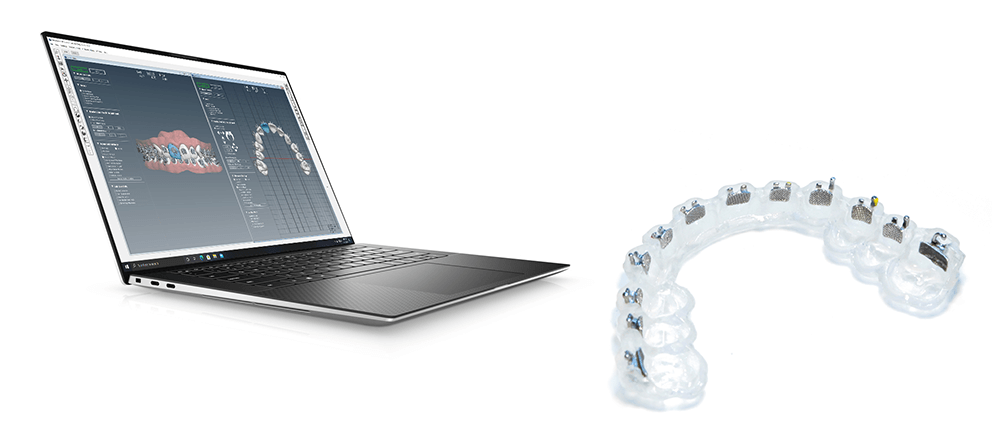
Digitized images of each patient’s teeth were created using a variety of commercially available intraoral scanners. These images were then saved as STL files and downloaded into the DIBS software to create digital models of the respective dentition. The software algorithms were then used to create desired virtual bracket positions on each of the teeth to be treated. In seven of the cases, individual orthodontists opted to just place brackets on one arch rather than both.
After each digital case setup was completed and approved by the orthodontist, indirect bonding trays were 3D printed by OrthoSelect in the company facilities and loaded with the appropriate brackets for each patient. The trays were then returned to the orthodontists for placement on their patients’ teeth.
After the brackets were placed, light cured and trays removed, a second intraoral scan was completed on each patient to create a 3D model of the patients’ dentition with brackets in place. This second 3D in vivo model was then compared to the original digital model produced by the DIBS software using AutoCAD software from Autodesk Inc. to determine deviation in virtual vs actual bracket placement.
A total of 446 brackets were placed on patients’ teeth as part of this study. As such, 892 measurements were made (446 vertical and 446 horizontal) comparing predicted vs actual placement outcomes across the 23 patients and against the clinically accepted deviation range of 0.5 mm.
Results:
Based on the total of 446 bracket placements, the mean absolute horizontal deviation between predicted and actual bracket placement was 0.154 mm (median of 0.152 mm), with a standard deviation of 0.049 mm. Individual horizontal deviations ranged from 0.000 mm to 0.886 mm. For vertical placement deviation, the absolute vertical deviation mean was 0.087 mm (median of 0.086), with a standard deviation of 0.025 mm. Individual vertical deviations ranged from 0.001 mm to 0.627 mm. The overall mean deviation was 0.121 mm (median of 0.123), with a standard deviation of 0.033 mm (see Table 1 below for measurements by individual patient).

There were a few instances where the placement deviation of the bracket fell outside the clinically acceptable range of 0.5 mm. Of the 446 horizontal measurements, there were 11 (2.5%) that fell outside the acceptable range. For the 446 vertical measurements, there was only one (0.2%) that fell outside the acceptable range. For all horizontal and vertical measurements combined, the total of 12 resulted in 1.3% outside the acceptable range. Accordingly, of all 446 brackets placed, 97.3% were measured within the range of 0.5 mm deviation along both horizontal and vertical axes.

There were a few instances where the placement deviation of the bracket fell outside the clinically acceptable range of 0.5 mm. Of the 446 horizontal measurements, there were 11 (2.5%) that fell outside the acceptable range. For the 446 vertical measurements, there was only one (0.2%) that fell outside the acceptable range. For all horizontal and vertical measurements combined, the total of 12 resulted in 1.3% outside the acceptable range. Accordingly, of all 446 brackets placed, 97.3% were measured within the range of 0.5 mm deviation along both horizontal and vertical axes.
Conclusions:
The results of this study indicate a high level of precision for bracket placement based on deviation between digitally modeled placement and actual bracket placement on patients’ dentition in both horizontal and vertical axes across several orthodontic practices and multiple patients. The overall mean deviation between modeled and actual bracket placement of 0.121 mm is well within the clinically accepted range of 0.5 mm.
In the 12 instances where bracket placement fell outside the acceptable range of 0.5 mm, several possible explanations may apply:
- Potential deliberate movement of the bracket by the orthodontist after placement and tray removal
- The quality of the post-placement scan of the bracket may have affected the results of the measurement
- Given that the majority of the larger variances were clustered in just a few patients, there may also have been some idiosyncratic factors at play (e.g., patient cooperation, practitioner clinical technique, etc.)
While larger variances certainly warrant further investigation, the overall low misplaced bracket incidence rate of only 2.7% in this study does not appear to indicate a systemic issue.
Overall, based on the data from this study, the DIBS platform (including digital modeling, transfer trays and bracket bonding) appears to provide a highly accurate protocol for bonding brackets to patients’ dentition.
1 Menini, A., Cozzani, M., Sfondrini, M.F. et al. A 15-month evaluation of bond failures of orthodontic brackets bonded with direct versus indirect bonding technique: a clinical trial. Prog Orthod 2014; 15:70.
2 Joiner M. In-house precision bracket placement with the indirect bonding technique. Am J Orthod Dentofacial Orthop 2010; 137:850-54.
3 Xue C, Xu H, Guo Y, Dhami Y, Wang H, Liu Z, Ma J, Bai D. Accurate bracket placement using a computer-aided design and computer-aided manufacturing-guided bonding device: An in vivo study. Am J Orthod Dentofacial Orthop. 2020; 157:269-77.